Interlaminar glia in health and disease
by Jorge A Colombo
Astroglial participation in the regulation of ionic balance, energy metabolism, morphogenesis, and neuritogenesis and synthesis of trophic factors, as well as synthesis and metabolism of neurotransmitter amino acids, confirms glial participation in multiple aspects of brain organization and function.
Although Andriezen in 1886 described glial cells with long processes in the human cerebral cortex, full morphological, developmental, and species comparative analysis was not attained until several years later. Probably due to prevalent rodent brain analysis is taken as a model to generalize their role in mammalian brain, histological, physiological, comparative, and pathological concepts -as well as their emergence during human brain development- regarding cerebral cortex astroglia were not systematically analyzed until several years later.
Health
Albeit with variations in the density of Glia with interlaminar processes, they are present in the cerebral cortex of humans and New and Old-World monkeys, but not in the rodent (Figure 1). We proposed to label such glial morphotype as interlaminar glia due to their processes (IGP) traversing several cellular layers of the cerebral cortex.
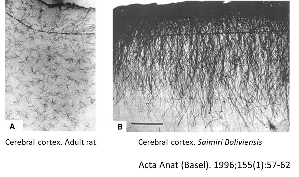
Although the functional significance of interlaminar astroglial processes is largely unknown, it has been speculated that they represent unusually long-ranging astroglial structures that may be responsible for remote, radially organized (columnar associated) and glial-mediated influences on the neurochemical environment in the cerebral cortex. Thus, interlaminar processes may provide a different spatial configuration and dynamics for neuronal-glial interaction in the cerebral cortex, as compared to the one resulting from the conventional stellate astrocyte (Colombo et al., Anat. Embryol. 1998).
Long, glial fibrillary acid protein (GFAP)-immunoreactive (IR) astroglial processes forming ordered palisades have been characterized as a specialized feature of the primate cerebral cortex, and are most highly developed in anthropoid species (Colombo et al.,1997; Colombo et al., 1998; Colombo et al., 2000). We have previously termed these structures as “interlaminar processes” because they span several cortical layers. These specialized astrocytic processes develop postnatally [and extend downward from superficial astrocytic cell bodies, traversing the supragranular layers in a predominantly radial fashion (Colombo et al., 2002).
Alzheimer Disease
In most cortical regions of cases diagnosed as severe Alzheimer’s disease by the donor institutions, interlaminar astroglia was found to be markedly altered or absent, and replaced by hypertrophic intralaminar astrocytes.
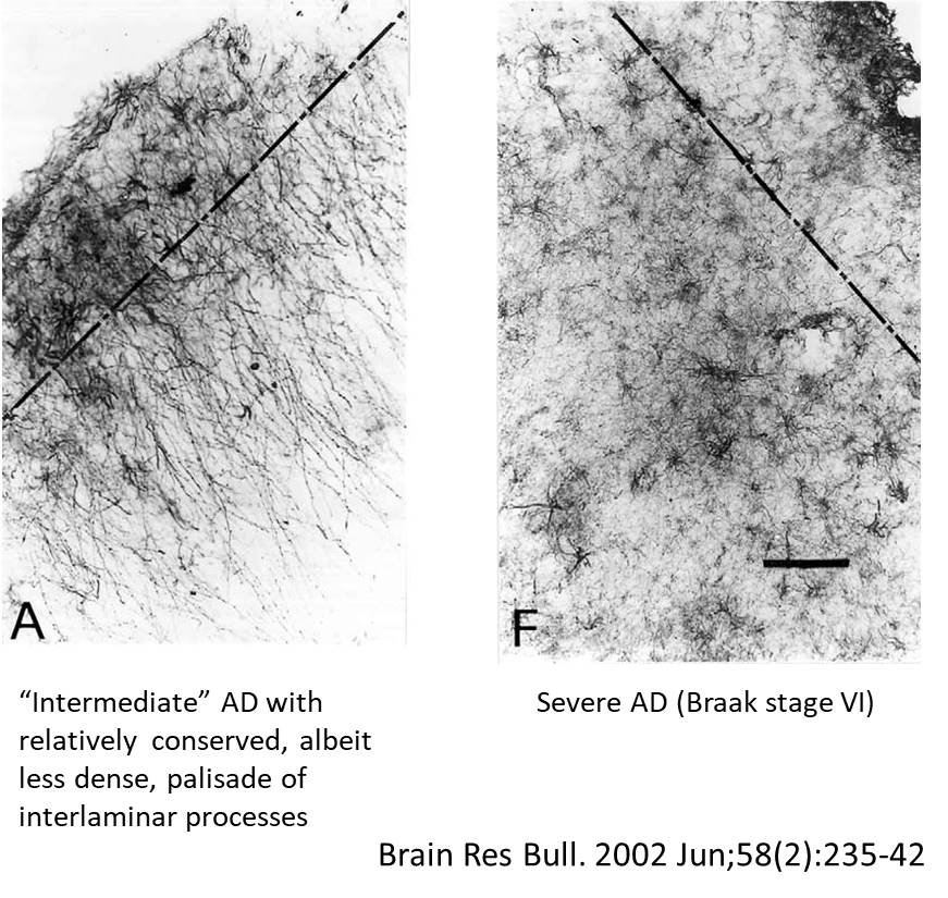
In general, the present observations suggest that the interlaminar processes are rather labile structures compared to intralaminar processes, that they do not form part of the chronic astrocytic reactive condition, and that they may be highly dynamic structures undergoing remodeling processes upon signals from the neuropil.
Down’s Syndrome
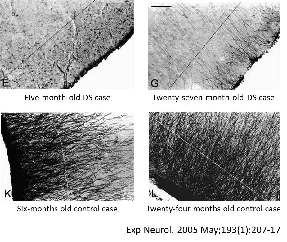
Comparing cerebral cortex control infant cases with age matched cases with Down’s Syndrome cases, the initial organization of the IGP was similar in control and DS cases, although a breakdown in DS became manifest by the first year of age, or earlier, albeit with individual variations. These changes tended to evolve in a mosaic fashion and included partial disruption of the palisade, or persistence of the physiological astrocytosis.
As proposed earlier, the evolution of the ‘‘primate-like’’ Interlaminar Glial Palisade (IGP) in the cerebral cortex may be schematically viewed as follows: bulk of non-primate species (lack of interlaminar processes) –Chiroptera and Insectivora (scanty, isolated IGP, limited to allocortex) –Strepsirrhini (lemuriforms) (variable presence of IGP)– Haplorrhini [Platirrhini (New World\Catarrhine (Old World) (in general well-developed IGP palisade, although variable in New World species)–anthropoid species (great apes, Homo) (well-developed IGP palisade).
Comments
On speculative grounds, the presence of IGP predominant in anthropoid species could be associated with optimizing neuronal columnar organization of the primate cerebral cortex. Their progressive installation during development is probably associated with maturation of brain function. Their disruption in Alzheimer’s Disease and Down’s Syndrome cases would correlate with brain progressive dysfunction.
References
Andriezen WL. (1893) The neuroglia elements of the brain. Brit Med J 29:227–230.
Colombo JA, Hartig W, Lipina S, Bons N. (1998) Astroglial interlaminar processes in the cerebral cortex of prosimians and Old-World monkeys. Anat Embryol 197:369–376.
Colombo JA, Quinn B, Puissant V. (2002) Disruption of astroglial interlaminar processes in Alzheimer’s disease. Brain Res Bull 58:235–242.
Colombo JA, Reisin HD, Jones M, Bentham C. (2005) Development of interlaminar astroglial processes in the cerebral cortex of control and Down’s syndrome human cases. Exp Neurol 193:207–217.
Colombo JA.; Yáñez A; Lipina S (1997) Interlaminar astroglial processes in the cerebral cortex of nonhuman primates: Response to injury. J. Brain Res. 38:503–512.
Colombo, JA; Fuchs E.; Hartig W.; Marotte L.; Puissant V. (2000) Rodent-like and primate-like types of astroglial architecture in the adult cerebral cortex of mammals. A comparative study. Anat. Embryol. 201:111–120.
Author: Jorge A. Colombo MD, PhD
Principal Investigator (CONICET) (retired)
Buenos Aires
Argentina
-COLOMBO J.A.The interlaminar glia: from serendipity to hypothesis 2017. BRAIN STRUCTURE AND FUNCTION. 10.1007_s00429-016-1332-8 (2).pdf
Suggested further reading on this subject:
-COLOMBO J.A.The interlaminar glia: from serendipity to hypothesis 2017 10.1007_s00429-016-1332-8 (2).pdf
-COLOMBO J.A.Interlaminar Glia and Other Glial Themes Revisited: Pending Answers Following Three Decades of Glial Research. Neuroglia 2018, 1, 3; doi:10.3390/neuroglia1010003